Reisenauer, H. P.; Wagner, J. P.; Schreiner, P. R. Angew. Chem. Int. Ed. 2014, 53, 11766-11771
Contributed by Steven Bachrach.
Reposted from Computational Organic Chemistry with permission


This work is licensed under a Creative Commons Attribution-NoDerivs 3.0 Unported License.
Contributed by Steven Bachrach.
Reposted from Computational Organic Chemistry with permission
I remain amazed at how regularly I read reports of structure determinations of what seem to be simple molecules, yet these structures have eluded determination for decades if not centuries. An example is the recently determined x-ray crystal structure of L-phenylalanine;1 who knew that growing these crystals would be so difficult?
The paper I want to discuss here is on the gas-phase structure of carbonic acid 1.2 Who would have thought that preparing a pure gas-phase sample would be so difficult? Schreiner and co-workers prepared carbonic acid by high-vacuum flash pyrolysis (HVFP) of di-tert-butyl carbonate, as shown in Scheme 1.
Scheme 1

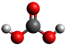
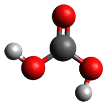
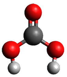
Carbonic acid can appear in three difference conformations, shown in Figure 1. The two lowest energy conformations are separated by a barrier of 9.5 kcal mol-1 (estimated by focal point energy analysis). These conformations can be interconverted using near IR light. The third conformation is energetically inaccessible.
1cc
(0.0) |
1ct
(1.6) |
1tt
(10.1) |
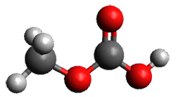
2cc
|
2cc
| |
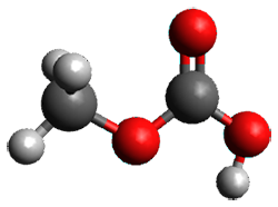
Figure 1. CCSD(T)/cc-pVQZ optimized structures of 1 (and the focal point relative energies in kcal mol-1) and the CCSD(T)/cc-pVTZ optimized structures of 2.
The structures of these two lowest energy conformations were confirmed by comparing their experimental IR spectra with the computed spectra (CCSD(T)/cc-pVTZ) and their experimental and computed rotational constants.
An interesting added component of this paper is that sublimation of the α- and β-polymorphs of carbonic acid do not produce the same compound. Sublimation of the β-isomorph does produce 1, but sublimation of the α-isomorph produces the methylester of 1, compound 2 (see Figure 1). The structure of 2 is again confirmed by comparison of the experimental and computed IR spectra.
References
(1) Ihlefeldt, F. S.; Pettersen, F. B.; von Bonin, A.; Zawadzka, M.; Görbitz, C. H. "The Polymorphs of L-Phenylalanine," Angew. Chem. Int. Ed. 2014, 53, 13600–13604, DOI: 10.1002/anie.201406886.
(2) Reisenauer, H. P.; Wagner, J. P.; Schreiner, P. R. "Gas-Phase Preparation of Carbonic Acid and Its Monomethyl Ester," Angew. Chem. Int. Ed. 2014, 53, 11766-11771, DOI: 10.1002/anie.201406969.
InChIs
1: InChI=1S/CH2O3/c2-1(3)4/h(H2,2,3,4)
InChIKey=BVKZGUZCCUSVTD-UHFFFAOYSA-N
InChIKey=BVKZGUZCCUSVTD-UHFFFAOYSA-N
2: InChI=1S/C2H4O3/c1-5-2(3)4/h1H3,(H,3,4)
InChIKey=CXHHBNMLPJOKQD-UHFFFAOYSA-N
InChIKey=CXHHBNMLPJOKQD-UHFFFAOYSA-N

This work is licensed under a Creative Commons Attribution-NoDerivs 3.0 Unported License.
No comments:
Post a Comment